Welcome to the Alexander Yule Consulting Blog

Showing posts with label microbiome. Show all posts

Thursday, 29 November 2018

Linking the brain, gut and bacteria in neurological disease.


The lifetime risk of developing neurological disease is influenced by variety of factors: genetics, cardiovascular health, and of course, age and neuroscience research continues to uncover more subtle links. 

Recent work elaborates on a long-suspected connection between that occasionally troublesome leftover, the appendix, and Parkinson’s disease risk, while other researchers have raised the possibility of the brain having its own microbiome, with implications for a bacterial influence on the risk and course of neurological disease.

Alzheimer’s disease (AD) and Parkinson’s disease (PD) are characterised by the accumulation of mis-folded proteins in the brain: amyloid β and tau proteins in the case of AD, and α-synuclein in PD, where it is the main constituent of “Lewy bodies”- clumps of aggregated protein found within neurons and a hallmark of PD and other dementias. Mutations within the α-synuclein gene are found in familial PD and efforts are ongoing to determine the normal function of α-synuclein and whether preventing its aggregation or accumulation in the brain might be of benefit.

PD causes both motor and non-motor symptoms and gut-associated problems such as constipation and impaired emptying of the stomach are common years before the onset of motor symptoms. That the aberrant form of α-synuclein can be found throughout the gastrointestinal tract in individuals with PD has been known for several years, although this also the case in those who don’t develop PD.
   
The highest levels of aberrant α-synuclein are found the appendix, raising the possibility that it serves as reservoir for dysfunctional protein which makes its way to the brain via the vagus nerve and potentiates the transformation of normal α-synuclein into the aggregated form. Circumstantial evidence for a link between the appendix and PD risk has been found in a large epidemiological study, with individuals who had undergone surgical severing of the vagus nerve (usually to manage hard to treat chronic duodenal ulcers) being at  lower risk of developing PD.
  
Defining the role of the appendix in PD has proved elusive, and three recent epidemiological studies failed to find any obvious link. By analysing medical records from over 1.6m Swedes from 1964, a research group at the Van Andel Institute has established that appendectomy reduces the risk of developing PD by around 20%, although this protective effect was only apparent in individuals living in rural areas. Pesticide and herbicide exposure are linked with a higher risk of PD and appendectomy may in some way mitigate environment-related PD risk.  Further analysis indicated that appendectomy delayed the onset of PD by an average of more than three and a half years in those who had undergone appendix removal 30 years or more before.

Biochemical analysis of appendix samples from healthy individuals and those with PD identified aberrant forms of α-synuclein. These were present in 46 of 48 normal individuals.  Mixing normal appendix tissue with normal α-synuclein resulted in the protein being broken down into forms resembling those found in PD brain samples.
 
Although far from being a recommendation for elective appendectomy, the finding that aberrant α-synuclein is common in healthy people suggests that PD risk requires its migration to the brain. Finding ways of confining, or even eliminating, the protein from the appendix could conceivably reduce PD risk.

It’s postulated that appendix might play a role in monitoring and restoring the gut microbiome and that inflammation results in changes which favour bacteria which generate “pro-PD” metabolites. That the gut flora might directly influence the neurological environment is not as far-fetched as it once would have seemed although a poster presentation given at the Society for Neuroscience annual meeting suggests the possibility that a local, rather than distant, microbiome might potentially influence conditions in the brain.

University of Alabama researchers have found that rod-shaped structures first observed on electron microscopic examination of brain samples from schizophrenics are, in fact, bacteria. These were most abundant in the substantia nigra, hippocampus and prefrontal cortex but rarer in the striatum. Bacteria were also found within brain cells, particularly in the ends of astrocytes closest to the blood-brain barrier locations, in dendrites, glial cells and in and around myelinated axons.

To rule out sample contamination, the group compared fresh brain samples from mice raised in a normal environment and those born and maintained in a germ-free environment: bacteria were only found in the former. Nucleic acid sequencing indicated that most of the bacteria belonged to groups commonly found in the gut, although their means of passage to the brain- whether from blood, the nose or through the nervous system.

Since bacteria were found in the brains of both normal individuals and those with schizophrenia, there’s no obvious causal relationship, but, as the study of gut, oral and skin microbiomes has shown, bacterial nutrients and metabolites can cause subtle but important changes in cell and organ function. Whether the presence of bacteria in the brain truly indicates a permanent ecosystem and not merely a post-mortem artefact remains to be established. But, as with the appendix and PD, confirmation that the brain is indeed influenced by local (or distant) bacteria may help better define neurological disease risk and uncover new means of treatment and prevention.

Tuesday, 26 June 2018

Bugs as drugs?

Lactococcus lactis: Potential
beyond cheesemaking? 
More microbiological nostalgia, this time prompted by an article in Nature concerning another decades-old concept, the therapeutic use of genetically engineered bacteria.

The early 90s saw a spate of clinical studies involving bacteria (Clostridium and Salmonella species) capable of growing and multiplying within the oxygen-poor environment found within solid tumours, their natural anti-cancer properties enhanced through engineered expression of enzymes capable of activating cancer drugs. Response rate were far from compelling, although interest remains in their use as oncolytic “vaccines".

Listeria monocytogenes is under evaluation as a live vector for the delivery of tumour-specific antigens across a variety of cancer indications, but the treatment-related death of a cervical cancer patient (and subsequent clinical hold) in a Phase I/II combination study involving Advaxis’s L.monocytogenes HPV vaccine has raised questions as to whether the vector or AstraZeneca’s immune checkpoint, Imfinzi® (durvalumab) is the culprit.

More recent efforts are directed towards conditions other than cancer and are exploiting advances made in synthetic biology. A US company, Synlogic, is applying “therapeutic programming” to repurpose commensal bacteria by stitching in DNA encoding for environmental sensors and metabolic switches. Clinical studies have recently commenced with an E.coli strain engineered to break down phenylalanine, an amino acid that accumulates to harmful effect in those with the hereditary condition phenylketonuria, and with another engineered strain capable of breaking down ammonia in those with life-threatening blood levels arising from genetic disease or through liver failure.

Intrexon (through a subsidiary, ActoBio Therapeutics and various collaborators) is looking to repurpose the cheese makers’ friend, Lactobacillus lactis. A clinical study is underway with a strain that produces trefoil factor I, a human protein involved in the maintenance and repair of mucosal epithelium, in subjects with oral mucositis, a painful inflammation of the lining of the mouth and a common side effect of radio- and chemotherapy.

Another strain in the clinic produces an antibody fragment against tumour necrosis factor, the target of several established and effective therapies for inflammatory bowel disease and a study is planned with a strain which produces a form of insulin thought to trigger the autoimmune destruction of insulin-secreting cells in Type 1 diabetes, along with a tolerance-inducing cytokine.

While the genetic manipulation of well-characterised bacteria is relatively straightforward, the design and development of effective therapeutic strains is not without problems. Mutation and the loss of inserted plasmids can result in reversion of engineered bacteria to their wild state. Predicting the potency of modified bacteria and the likelihood of successful colonisation remains complex and uncertain. As demonstrated by antibiotic resistance, bacteria are notoriously promiscuous when it comes to sharing DNA and there is a risk that the ability to express therapeutic proteins might be passed to other bacterial species. The gut and oral microbiomes play subtle and important roles in human health and might be detrimentally altered through colonisation by engineered strains.

While none of these challenges are insurmountable, and with admiration at the ingenuity exercised in designing and developing bacterial therapeutics, I suspect that the same treatment objectives can be more readily achieved by established, less problematic, technologies, although the convergence of synthetic biology and growing understanding of the human microbiome opens some intriguing, if not near-horizon, possibilities for short or long term beneficial manipulation.

Photo credit: Kenneth Toda, University of Wisconsin

Saturday, 3 March 2018

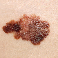
Another “bugs and cancer” post. Can bacteria reduce the risk of skin cancer?

Melanoma lesion. Do bacteria contribute to 
our skin cancer defences?
Research into (and clinical and commercial exploitation of) our bacterial travelling companions has so far largely focused on the relationship between gut dwellers and health. As the largest organ of the body (at between 6% and 10% of total body weight), and with the function of keeping the inside safe from the outside, it’s perhaps not surprising that human skin has evolved a rich and complex microbiome which can influence the course of disease.

In common with Planet Earth as described in the “Hitchhiker’s Guide to the Galaxy”, skin bacteria are “mostly harmless”, although physiological changes can make us susceptible to blemishes and rashes caused largely by Staphylococcus aureus and, that bane of teenagers, Propionibacterium acnesSkin bacteria in the wrong place (the blood stream, newly-replaced hip joints) are always bad news.

But, by and large, the skin microbiome functions well to keep potential pathogens in check through a variety of mechanisms, including production of selective antibacterial agents and the downregulation of inflammation. One of the most studied of these “good guy “bacteria is Staphylococcus epidermis. Collaboration between researchers at the University of California, San Diego, and other institutes has uncovered a possible association between certain strains of S.epidermis and suppression of the skin cancer, melanoma.

While attempting to better characterise the selective antibacterial activity of S.epidermis, the UCSD group identified a strain producing 6-N-hydroxyaminopurine (6-HAP), a suppressor of DNA synthesis. S.epidermis 6-HAP was established to be non-toxic, with no evidence of the mutagenic effects associated with chemically-synthesised 6-HAP. When tested in the laboratory, 6-HAP was active against squamous cell carcinoma and melanoma cell lines, and to a lesser extent, lymphoma cells. 

Systemic treatment of mice in which human melanoma tumours had been established with 6-HAP resulted in a 60% reduction in tumour size, while colonization of the skin of mice with the 6-HAP-producing S.epidermis strain significantly reduced the occurrence of UV light-induced skin cancer over mice identically treated with a non-6-HAP-producing strain.

Early days, but this does raise the possibility that in addition to their established “anti-pathogen” role, S.epidermis and possibly other commensal skin bacteria might actively contribute to tumour suppression through early elimination of transformed cells. The 6-HPA-producing  strain appears to be common “in the wild” and epidemiological studies might perhaps someday establish an association between 6-HPA producers (or their absence) and melanoma prevalence. 

The UCSD researchers are hopeful of exploiting their findings through a start-up company, MatriSys Bioscience, with the notion of restoring or modifying the skin microbiome. Effecting colonization with a 6-HAP-producing S.epidermis strain could conceivably reduce susceptibility to melanoma and other skin cancers, but given the multifactorial nature of cancer, it’s not something that will be easy to establish in the context of clinical studies. Nevertheless, the UCSD study does underscore that outer microbiome research has an importance beyond better treatments for eczema and acne.

Photo credit: NCI

Monday, 11 December 2017

Yet more on bugs and cancer

T-cells (red) on the attack
A Research Highlights piece in December’s Nature Reviews Cancer reports on another intriguing aspect of the interplay between our immune systems and the bugs we carry, namely how gut flora might influence the effectiveness of cancer immunotherapy.

Two international research groups set out to determine  whether the composition of the gut microbiome might influence the response to immunotherapy  directed against  PD-1, a so-called “immune checkpoint “ expressed by activated T cells and macrophages and which is exploited by cancer cells to switch off immune attack. Antibody-mediated blockade of the interaction between PD-1 and its ligand, PD-L1 can restore the anti-cancer response. The anti-PD-1 antibodies pembrolizumab and nivolumab (Opdivo® and Keytruda®, respectively) have proved their worth in the treatment of metastatic melanoma and a variety of other solid tumours.

Genetic analysis of faecal bacteria collected from cancer patients before and after anti-PD-1 immunotherapy found a correlation between gut bacteria diversity and the duration of progression-free survival in cancer patient after treatment.

A collaboration between US and French researchers found differences in the abundance of certain gut bacteria, with Faecalibacterium being enriched in melanoma patients responsive to antiPD1 therapy: Bacteroidales was enriched in those patients not responsive to immunotherapy. Differences were also found between responders and non-responders in regards to bacterial metabolism and the composition of immune cells found in the tumour microenvironment. Tumour-infiltrating “killer” T cells were more likely to be found in patients carrying an abundance of Faecalibacterium, while  immunosuppresive cells were more common in individuals carrying abundant Bacteroidales.

Another (again, predominantly American and French) research group found that the abundance of the gut bacterium Akkermansia muciniphila in non-small cell lung cancer and renal cancer patients correlated with a positive response to anti-PD-1 immunotherapy.

Both groups looked for possible mechanistic links between gut bacteria abundance and treatment response. When patient-derived gut bacteria were transplanted into germ-free mice, a variety of favourable effects on tumour growth and immune response were observed, including higher numbers of killer T-cells  and other, immune effector cells, along with changes in the expression of  T- cell receptors for key immune signalling molecules (“chemokines”).

The response to immunotherapy is difficult to predict and involves a variety of tumour factors (PD-L1 expression, tumour burden, degree of mutation) and host factors (immune system genetic makeup, T cell infiltration of the tumour). Analysis of the gut microbiome is unlikely to improve prediction of response, but preservation or manipulation of the gut microbiome through avoidance of antibiotic treatment prior to immunotherapy, or probiotic treatment to encourage “good” bacteria could conceivably translate into better and more sustainable response rates for at least some individuals.  

Photo credit : Rita Elena Serda.  National Cancer Institute \ Duncan Comprehensive Cancer Center at Baylor College of Medicine

Saturday, 2 December 2017

Bugs and cancer? The plot thickens...

An item from the New York Times gives me the chance to write about two great interests in the same blog piece: bacteria (once a microbiologist, always a microbiologist) and cancer.
Fusobacterium nucleatum, an
accomplice of colorectal cancer

That bacterial infection might cause or promote cancer was debated for most of the 20th century, but with little solid evidence emerging to support the notions. During the 1980s, Barry Marshall and Robin Warren (the former famously swigging down a flask of culture broth to prove his hypothesis) established that Helicobacter pylori, a common corkscrew-shaped found in the stomach, was an undisputable cause of gastric inflammation and ulcers.

Epidemiological studies involving British, American and Japanese subjects confirmed that H.pylori carriage was indeed associated with an almost four-fold increase in the likelihood of developing gastric cancer and resulted in the WHO designating H.pylori as a Class I carcinogen.

Continuing research has established that the relationship between  H.pylori and cancer is not a simple one of cause and effect,  with H.pylori infection being a factor in some, but not all, forms of stomach cancer and that H.pylori  strains expressing a particular cytotoxin, “CagA”,  are more strongly associated with an elevated risk of cancer than are non-producing strains. Perversely, H.pylori infection appears to be associated with a lower risk of oesophageal cancer.

A more recently uncovered “smoking gun” is the presence of Fusobacterium nucleatum, a common mouth-dweller, found in higher numbers in around half of colorectal tumours than in the surrounding tissue. F.nucleatum- induced inflammation is cited as a plausible contributor to CRC initiation and progression.

But, as with the Helicobacter story, there is no clear-cut cause and effect between infection and cancer. Bacterial species are rarely solitary and the inhabitants of the local milieu or “microbiome” may be more important with respect to cancer initiation and/or progression than the presence of F.nucleatum alone.

CRC may spread to other organs and give rise to tumours in the liver. According to a recent Science publication, if F.nucleatum and its microbiome buddies are present in the original tumour, then they can accompany the metastasizing cancer and pitch up in the liver. CRC dwelling F.nucleatum remained associated with tumours even after their transplantation into mice. Moreover, dosing of tumour-bearing mice with an F.nucleatum-killing antibiotic slowed tumour growth.

Does this make a case for antibiotic therapy or vaccine development to reduce CRC rates? Well, not yet. Antibiotics therapy tends to ablate both the good and bad and, as is hinted at in immuno-oncology studies, certain gut bacteria might positively influence anti-cancer immune responses. And not all F.nucleatum strains might be bad guys. However, it’s feasible that getting a better handle on the mechanism(s) involved in the bacterial promotion of cancer might identify new interventions to improve outcomes or recurrence rates.

Photo credit: CDC Public Image Library